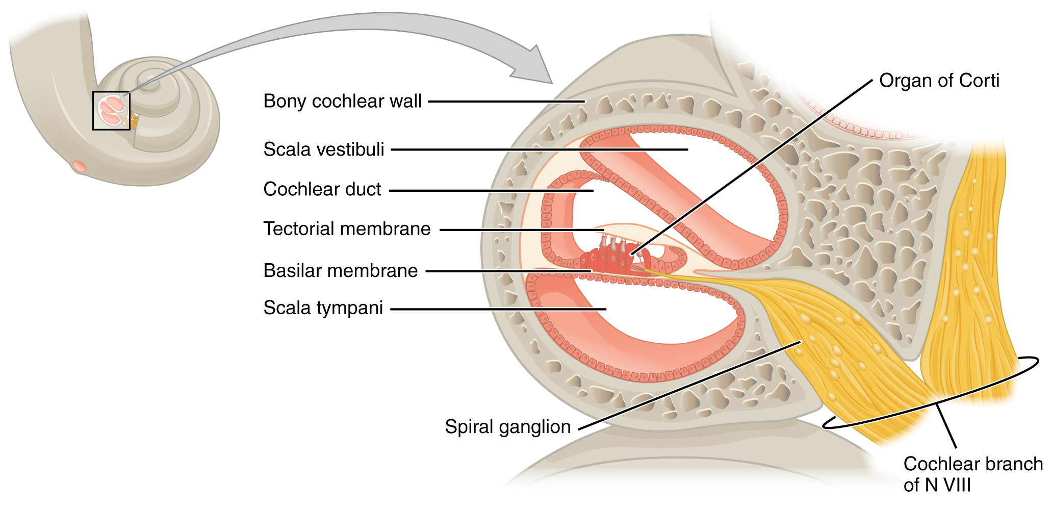
The cochlea is a spirally coiled tube (2 1/2 turns) of about an inch around a central, cone-slhaped bony pillar, the modiolus.
It lies deeply embedded in bone which resembles in shape to a snail’s shell. The cochlea consists of three tapering, parallel canals.
ADVERTISEMENTS:
The uppermost canal known as vestibular canal (scala vesti- buli) is attached at its end to the membrane-covered oval window (fenestra ovalis). This window holds the base of a small middle ear bone, the stapes.
At its other end located at the apex of the cochlea the vestibular canal has a small opening which communicates with the lowermost canal called the tympanic canal (scala tympani)?
The tympanic canal also has a membrane-covered round window (fenestra rotundum) at its base end, near the middle-ear chamber, but has no bone fitted to its surface.
Both the vestibular and tympanic canals are filled with a clear fluid called perilymph which comes from the cerebrospinal fluid and remain connected at the tip of the cochlea by a narrow passage called helicotrema.
ADVERTISEMENTS:
The third and the smallest central canal called the cochlear canal (scala media) is filled with a clear fluid called endolymph and remains separated from the overlying vestibular canal by a membrane called the vestibular membrane (Reissner’s membrane) and from underlying tympanic canal by a ledge-like projection of the bony cochlear wall plus a membrane, the basilar membrane.
The actual receptors for hearing are present in the cochlear canal as several rows of specialized “hair cells”, approximately 24,000 in number.
They contain numerous cilia (stereocilia) which project into the endolymph from the free end of each cell.
The rows of hair cells together with supporting cells and surrounding dendrites constitute the specialized organ of Corti.
ADVERTISEMENTS:
It rests on the basilar membrane within the cochlear canal. The cell bodies from which the dendrites originate make up a ganglion with one of the numerous passages of the inner ear; their axons form the auditory nerve a branch of one of the cranial nerves.
Sound is transmitted in the ear as follows. Vibrations enter the auditory canal and cause the tympanic membrane to vibrate.
Vibrations of the tympanic membrane are transmitted through middle-ear bones or ear ossicles to the membrane of fenestra ovalis or oval window, and then pass to the perilymph and scala vestibuli (vestibular canal) causing the Reissner’s (or vestibular) membrane to vibrate up and down, these vibrations pass to the endolymph which makes the basilar membrane to vibrate by which sensory hair cells of organ of Corti are stimulated, from here impulses travel through the fibres of auditory nerve to the brain where the temporal lobes of the cerebrum interpret them as sound.
Vibrations of the basilar membrane pass to the perilymph in scala tympani (tympanic canal) and the membrane of fenestra rotunda (round window) is bulged in and out to dampen the vibrations.
The helicotrema connecting the scala vestibuli (vestibular canal) with scala tympani (tympanic canal) is very narrow and is unable to transmit vibrations.
Thus the sense of hearing is confined to the cochlea. The explanation of hearing is based on evidenee but is primarily a theoretical one.
The three semicircular canals and their cristae detect changes in acceleration in any of the three planes of space.
The cristae also react to rapid changes of position and rotations, this is due to movements caused in the endolymph. The macula of sacculus is concerned with detection of gravitational stimuli.

