Important kinds of sense organs found in prawn are described below:
(i) Compound eyes:
At the base of rostrum a black, hemispherical movable stalked eye is found to be lodged in an orbital notch.
ADVERTISEMENTS:
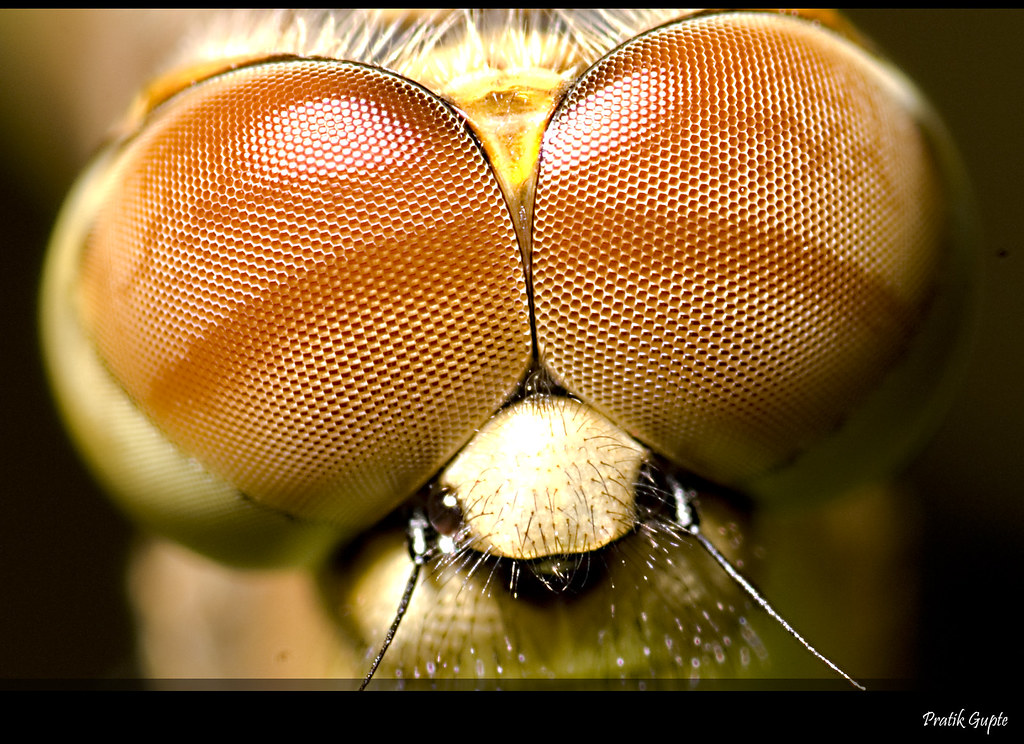
Each eye is compound eye and is made up of a large number (about 2500) of independent visual units, called ommatidia (Gr. Ommaton, little eye). This is the unique feature of phylum Arthropoda. All the ommatidia are radially arranged and structurally consist of two regions.
(A) Diopterical region:
This is outer region of the eye which focuses the light on receptor region of the eye. It has three layers, one below the other:
ADVERTISEMENTS:
Cornea:
This is the outermost layer of eye, which consists of transparent cuticle. Its surface has a large number of squares or facets with clearly visible lines and gives the appearance of a graph paper. Below such a facet lies one ommatidium.
Corneagen cells:
Each facet thickens in the centre to form a biconvex corneal lens. Beneath the lens lie two corneagen cells which secrete a new cornea as soon as the old one is cast off in moulting.
ADVERTISEMENTS:
Cone cells:
Beneath the corneagen cells lie four elongated cone cells or vitrellae which constitute a transparent, homogenous crystalline cone. Inner ends of cone cells are long and tapering.
(B) Receptor region:
This is a light sensitive inner region of eye. It consists of rhabdome and retinal cells :
Rhabdome and retinal cells:
Inner end of cone cells lie upon an elongated, spindle-shaped rod, the rhabdom. It is secreted and surrounded by a group of seven elongated retinal cells.
Retinal cells are layered upon basement membrane and are well connected with sensory nerve fibers of optic ganglia.
Chromatophores:
In between the ommatidium is a sheath of movable amoeboid dark pigment cells or chromatophores.
They are present in two layers, one outer layer is between the cone cells, called iris pigment and the inner one between the rhabdomes is called retinal pigment. Pigment cells vary in position according to the variation in the intensity of light.
Mosaic vision of the compound eye :
Working of compound eye is very complex. This is, however, deficient in focussing ability and clarity of image but efficient for picking up motion of the object and for peripheral vision.
It functions as a very efficient organ for photoreception. Movable stalked eyes provide 360-degree vision to the animal.
Each ommatidium makes separate image and all ommatidia combinedly make a mosaic image of an object because complete image is not single, instead it is total of all the separate small-small images formed from each separate ommatidium.
The nature of composite image formed varies according to different intensities of light. Two types of image are formed due to the movement of pigment cells.
(a) Apposition image:
This kind of image forms in bright light during day time. Pigment cells spread throughout along the ommatidia and separate them optically.
In this condition the rays of light which strike the cornea obliquely absorbed by the pigment cells. They do not reach the rhabdom and therefore produce no image.
Only those rays of light which fali perpendicularly upon the cornea can pass through the ommatidium and reach the rhabdom to form a point of image.
The other ommatidia form the images of other parts of the object. These all point images are fitted together into a single general picture.
It is known as mosaic or apposition image. Its sharpness depends upon the number of ommatidia involved and the degree of their isolation from one another.
(b) Superposition image:
In dim light (during night) the pigment cells migrate to be confined in distal and proximal segment. In this position the ommatidia are not optically separated but work in unison.
The oblique rays are not absorbed and are capable of forming a point of image after passing through a number of ommatidia instead of single ommatidia.
As a result, an overlapping of the adjacent point images occurs so that a continous or superposition image is obtained.
It is not sharp but the animal gets some sort of idea of the objects moving about in the surrounding.
The prawns, like most arthropods seem to adjust their eyes to form both types of images according to the prevailing intensity of light.
The optic nerve carries impulses (electrochemical waves of energy) to the brain, where they are interpreted and registered as an upright mental image,
(ii) Statocysts:
Statocysts are a pair of small white bead-like cuticular spherical sacs found inside the precoxa of each antennule.
It opens dorsally through a minute statocystic aperture, which remains covered by a small fold of integument.
It is supplied by a small statocystic branch of antennulary nerve. In transverse section the cavity of statocyst is oval, filled with minute sand particals and lined by a number of elongated delicate sensory hairs or receptor setae.
Each seta is supplied by a small branch of the statocystic nerve. Each setae has a swollen base towards the center.
Function:
Statocyst perceives the direction of the force of gravity and functions as the organ of orientation and equilibrium.
The sand particles function as statoliths. When position of palaemon changes in water, the sand grains press the setae and stimulate them.
The stimulated setae conveyed the massage to the brain so that the animal can correct its position.
(iii) Tangoreceptors:
Tangoreceptors are found in the form of plumose setae fringing the flat portions of appendages like the ramii of pleopods.
Each tactile seta is hollow cuticular outgrowth supplied with a nerve fiber. It consists of a swollen shaft and a blade.
The elongated feelers of both antennae are also said to be tactile in function.
(iv) Chemoreceptors:
Chemoreceptors or olfactory organs occur on mouthparts, flagella of antennules and inner wall of gill chambers. Each olfactory seta consists of basal shaft and a blade and innervated by a small nerve fiber from the olfactory branch.
