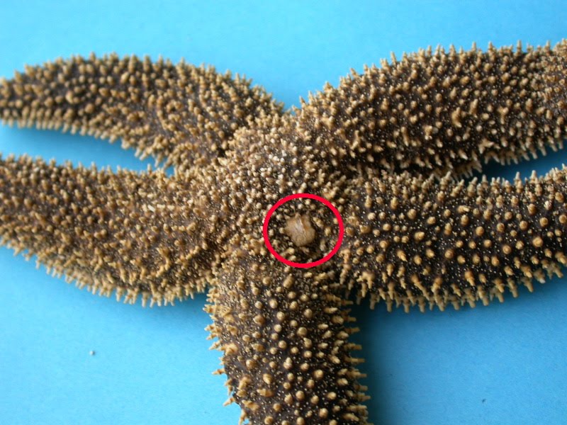
Important parts in the water vascular system or ambulacral system of asterias are listed below:
(1) Madreporite:
ADVERTISEMENTS:
It is a thick sieve like calcareous plate situated on the aboral surface of the central disc. It bears as many as 250 minute pores on the surface and they lead into pore canals.
All pore canals unite to form collecting canals within the substance of madreporite. Below the madreporite, collecting canal communicates with stone canal through ampulla.
(2) Stone canal:
It is S-shaped tube, which opens on the oral side into a ring canal around the mouth. It is also known as madreporite canal. Its walls are supported by a series of calcareous rings, hence the name stone canal. Inner lining of the wall bears cilia or flagella, which draw water into canal.
ADVERTISEMENTS:
Stone canal with an axial organ is enclosed in a coelomic sac, the axial sinus. These together form the axial complex.
(3) Ring canal:
It is a wide canal forming a ring around the oesophagus. The angles of the pentagonal ring lie in the radial positions.
(4) Tiedemann’s bodies:
ADVERTISEMENTS:
These are also known as racemose glands.They are small rounded yellowish glandular sacs opening into ring canal on its inner side.
There are total 9 tiedmann’s bodies arranged in radii and interradii positions except the position of stone canal.
It consists of a bounding peritonium enclosing a stroma of connective tissue and muscle fibers containing numerous radiating tubules.
The exact function of them is uncertain. Some workers consider them as filtering device, others as lymphatic glands which probably manufacture coelomocytes of water vascular system.
(5) Pollen vesicles:
These are pear-shaped, thin walled contractile bladders situated along the interradii and open into the ring canal on the outer side. They help in the regulation of pressure of sea water. In Asterias, however it is absent.
(6) Radial canals:
They arise from the ring canal and extend along each arm up to the tip. Radial canals lie below the ambulacral ossicles and terminate as the lumen of the terminal tentacles.
(7) Lateral canals:
Each radial canal in its corresponding arm gives out two series of narrow lateral or podial canals along its entire length.
Each lateral canal opens into a tube foot. The opening being provided by a valve to prevent back flow of fluid into radial canal
(8) Tube feet:
There are two double rows of tube feet in each arm. Each tube foot has the form of a close thin walled tube, distinguished into three regions-
(i) A rounded sac like ampulla situated above, the ambulacral ossicle and projecting into the coelom.
(ii) A middle tubular podium extending through the ambulacral groove and
(iii) A cup like sucker at the lower end of the podium.
Wall of the tube foot possesses strong longitudinal muscles. In addition, the walls of the ampulla also possess circular muscles while those of the podia possess rings of inelastic connective tissue.
Function:
Most peculiar and interesting role of water vascular system is in locomotion by providing a hydraulic pressure mechanism.
Thin walls of tube feet may serve for respiratory exchange of gases. Tube feet also help in anchoring the body to the substratum and in capturing and handling the food.

